Difference between revisions of "GCSE Biology Required Practical: Using a Microscope"
| (4 intermediate revisions by the same user not shown) | |||
| Line 1: | Line 1: | ||
==Key Stage 4== | ==Key Stage 4== | ||
| + | {{#ev:youtube|https://www.youtube.com/watch?v=SX6mow1AExI}} | ||
===Meaning=== | ===Meaning=== | ||
Use a [[microscope]] to make labelled [[diagram]]s of [[plant]] and [[animal]] [[Cell (Biology)|cells]] with a magnification scale. | Use a [[microscope]] to make labelled [[diagram]]s of [[plant]] and [[animal]] [[Cell (Biology)|cells]] with a magnification scale. | ||
| Line 5: | Line 6: | ||
===Method=== | ===Method=== | ||
====Observing Plant Cells==== | ====Observing Plant Cells==== | ||
| − | |||
| − | |||
| − | |||
{| class="wikitable" | {| class="wikitable" | ||
|- | |- | ||
| Line 13: | Line 11: | ||
|[[File:MicroscopePlantCells2.png|center|200px]] | |[[File:MicroscopePlantCells2.png|center|200px]] | ||
|[[File:MicroscopePlantCells3.png|center|200px]] | |[[File:MicroscopePlantCells3.png|center|200px]] | ||
| + | |- | ||
| + | | style="height:20px; width:200px; text-align:left;" |1. Place a [[sample]] of onion [[Plant Cell|cells]] on to a [[slide]]. | ||
| + | | style="height:20px; width:200px; text-align:center;" |2. Place a drop of 0.01M [[Iodine Solution]] onto the [[Cell (Biology)|cells]]. | ||
| + | | style="height:20px; width:200px; text-align:center;" |3. Place a cover slip on the [[Cell (Biology)|cells]] and use tissue paper to remove any excess [[Iodine Solution|iodine solution]] from the edges. | ||
|} | |} | ||
| + | : 4. Place the [[slide]] onto the [[stage]] of the [[microscope]] and focus the image. | ||
| + | : 5. Note the [[magnfication]] of the [[Eyepiece Lens|eyepiece lens]] and the [[Objective Lens|objective lens]]. | ||
| + | : 6. Sketch a [[Cell (Biology)|cell]] including a magnification scale to indicate how much larger you drawing is than the actual [[Cell (Biology)|cell]]. | ||
| − | |||
| − | |||
| − | |||
====Observing Animal Cells==== | ====Observing Animal Cells==== | ||
| − | |||
| − | |||
| − | |||
{| class="wikitable" | {| class="wikitable" | ||
|- | |- | ||
| Line 27: | Line 26: | ||
|[[File:MicroscopeAnimalCells2.png|center|200px]] | |[[File:MicroscopeAnimalCells2.png|center|200px]] | ||
|[[File:MicroscopeAnimalCells3.png|center|200px]] | |[[File:MicroscopeAnimalCells3.png|center|200px]] | ||
| + | |- | ||
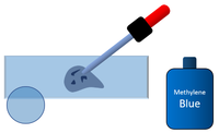
| + | | style="height:20px; width:200px; text-align:left;" |1. Place a [[sample]] of [[Animal Cell|animal cells]] on to a [[slide]]. | ||
| + | | style="height:20px; width:200px; text-align:center;" |2. Place a drop of [[Methylene Blue]] onto the [[Cell (Biology)|cells]]. | ||
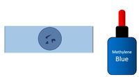
| + | | style="height:20px; width:200px; text-align:center;" |3. Place a cover slip on the [[Cell (Biology)|cells]] and use tissue paper to remove any excess [[Methylene Blue]] from the edges. | ||
|} | |} | ||
| − | + | :4. Place the [[slide]] onto the [[stage]] of the [[microscope]] and focus the image. | |
| − | + | :5. Note the [[magnfication]] of the [[Eyepiece Lens|eyepiece lens]] and the [[Objective Lens|objective lens]]. | |
| − | + | :6. Sketch a [[Cell (Biology)|cell]] including a magnification scale to indicate how much larger you drawing is than the actual [[Cell (Biology)|cell]]. | |
| + | ===References=== | ||
| + | ====Edexcel==== | ||
| + | :[https://www.amazon.co.uk/gp/product/1292120193/ref=as_li_tl?ie=UTF8&camp=1634&creative=6738&creativeASIN=1292120193&linkCode=as2&tag=nrjc-21&linkId=572df39392fb4200db8391d98ae6314e ''Biology core practicals; using microscopes, pages 6-7, GCSE Combined Science, Pearson Edexcel ''] | ||
Latest revision as of 14:27, 2 November 2019
Contents
Key Stage 4
Meaning
Use a microscope to make labelled diagrams of plant and animal cells with a magnification scale.
Method
Observing Plant Cells
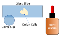
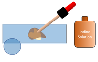
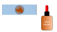
| 1. Place a sample of onion cells on to a slide. | 2. Place a drop of 0.01M Iodine Solution onto the cells. | 3. Place a cover slip on the cells and use tissue paper to remove any excess iodine solution from the edges. |
- 4. Place the slide onto the stage of the microscope and focus the image.
- 5. Note the magnfication of the eyepiece lens and the objective lens.
- 6. Sketch a cell including a magnification scale to indicate how much larger you drawing is than the actual cell.
Observing Animal Cells
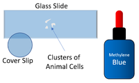
| 1. Place a sample of animal cells on to a slide. | 2. Place a drop of Methylene Blue onto the cells. | 3. Place a cover slip on the cells and use tissue paper to remove any excess Methylene Blue from the edges. |
- 4. Place the slide onto the stage of the microscope and focus the image.
- 5. Note the magnfication of the eyepiece lens and the objective lens.
- 6. Sketch a cell including a magnification scale to indicate how much larger you drawing is than the actual cell.
