Difference between revisions of "Trachea"
(→Key Stage 4) |
|||
| Line 15: | Line 15: | ||
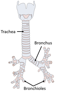
[[File:Trachea.png|right|200px|thumb|A diagram showing the [[trachea]].]] | [[File:Trachea.png|right|200px|thumb|A diagram showing the [[trachea]].]] | ||
The [[trachea]] is a [[cartilage]] covered tube connecting the [[mouth]] to the [[Lung|lungs]]. | The [[trachea]] is a [[cartilage]] covered tube connecting the [[mouth]] to the [[Lung|lungs]]. | ||
| − | |||
| − | |||
===Adaptations of the Trachea=== | ===Adaptations of the Trachea=== | ||
: The [[trachea]] is covered in [[cartilage]] to stop it from closing when the lungs take in [[Oxygen]]. | : The [[trachea]] is covered in [[cartilage]] to stop it from closing when the lungs take in [[Oxygen]]. | ||
Revision as of 09:35, 6 June 2019
Contents
Key Stage 3
Meaning

A diagram showing the trachea.
The trachea is a tube connecting the mouth to the lungs.
Adaptations of the Trachea
- The trachea is covered in cartilage to stop it from closing when the lungs take in Oxygen.
- The trachea contains specialsed cells which release mucus in order to trap micro-organisms and dust to prevent them entering the lungs.
- The inner lining of the trachea is covered in ciliated epithelial cells to sweep the mucus up away from the lungs.
About the Trachea
Key Stage 4
Meaning

A diagram showing the trachea.
The trachea is a cartilage covered tube connecting the mouth to the lungs.
Adaptations of the Trachea
- The trachea is covered in cartilage to stop it from closing when the lungs take in Oxygen.
- The trachea contains specialsed cells which release mucus in order to trap micro-organisms and dust to prevent them entering the lungs.
- The inner lining of the trachea is covered in ciliated epithelial cells to sweep the mucus up away from the lungs.